
Automated three-dimensional distance and coverage mapping of hallux valgus: a case-control study
DOI:
https://doi.org/10.30795/jfootankle.2022.v16.1629Keywords:
Imaging, three-dimensional, Hallux valgus, Metatarsal bones, Weight-bearingAbstract
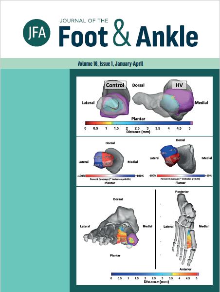
Objective: To develop distance-mapping and coverage-mapping algorithms to assess metatarsophalangeal and metatarsal-sesamoid joint interaction in hallux valgus patients, comparing them to a control group. Methods: A total of 9 hallux valgus patients (mean age 37.1 y; 6 F/3 M) and 5 controls (mean age 39 y; 4 F/1 M) underwent weight-bearing computed tomography. Specific software was used to obtain bone segmentation images of the first and second metatarsals, the first and second proximal phalanxes, and the tibial and fibular sesamoids. Joint interaction based on distance mapping and coverage mapping of the first and second metatarsophalangeal joints and the metatarsal-sesamoid joints were calculated. The surfaces of the metatarsophalangeal joints were divided in a 2-by-2 grid using the principal axes to provide a more detailed analysis. P-values <0.05 were considered significant. Results: Coverage maps of hallux valgus and control patients revealed marked lateral and dorsal displacement in joint interaction of the first metatarsophalangeal joint, including decreased joint coverage of the medial facet of the joint. When comparing first metatarsophalangeal joint coverage, hallux valgus patients had significantly lower coverage of the dorsomedial quadrant (77%, p=0.0002) than controls, as well as significantly higher coverage of the plantarlateral (182%, p=0.005) and dorsolateral quadrants (44.9%, p=0.035). Conclusions: In this case-control study, we developed a distance and coverage map weight-bearing computed tomography algorithm to objectively assess 3D joint interaction, joint coverage, and subluxation in hallux valgus deformity. We observed significantly greater first and second metatarsophalangeal joint subluxation in hallux valgus patients than controls. Level of Evidence III; Case Control Study.
Downloads
Published
How to Cite
Issue
Section
License
Copyright (c) 2022 Journal of the Foot & Ankle

This work is licensed under a Creative Commons Attribution-NonCommercial 4.0 International License.


