
Flexor hallucis longus. A cadaveric study of its distal insertion
DOI:
https://doi.org/10.30795/jfootankle.2023.v17.1693Keywords:
FHL, flexor hallucis longus, Hallux valgus, cadavericAbstract
Objective: Describe flexor hallucis longus (FHL) distal insertion.
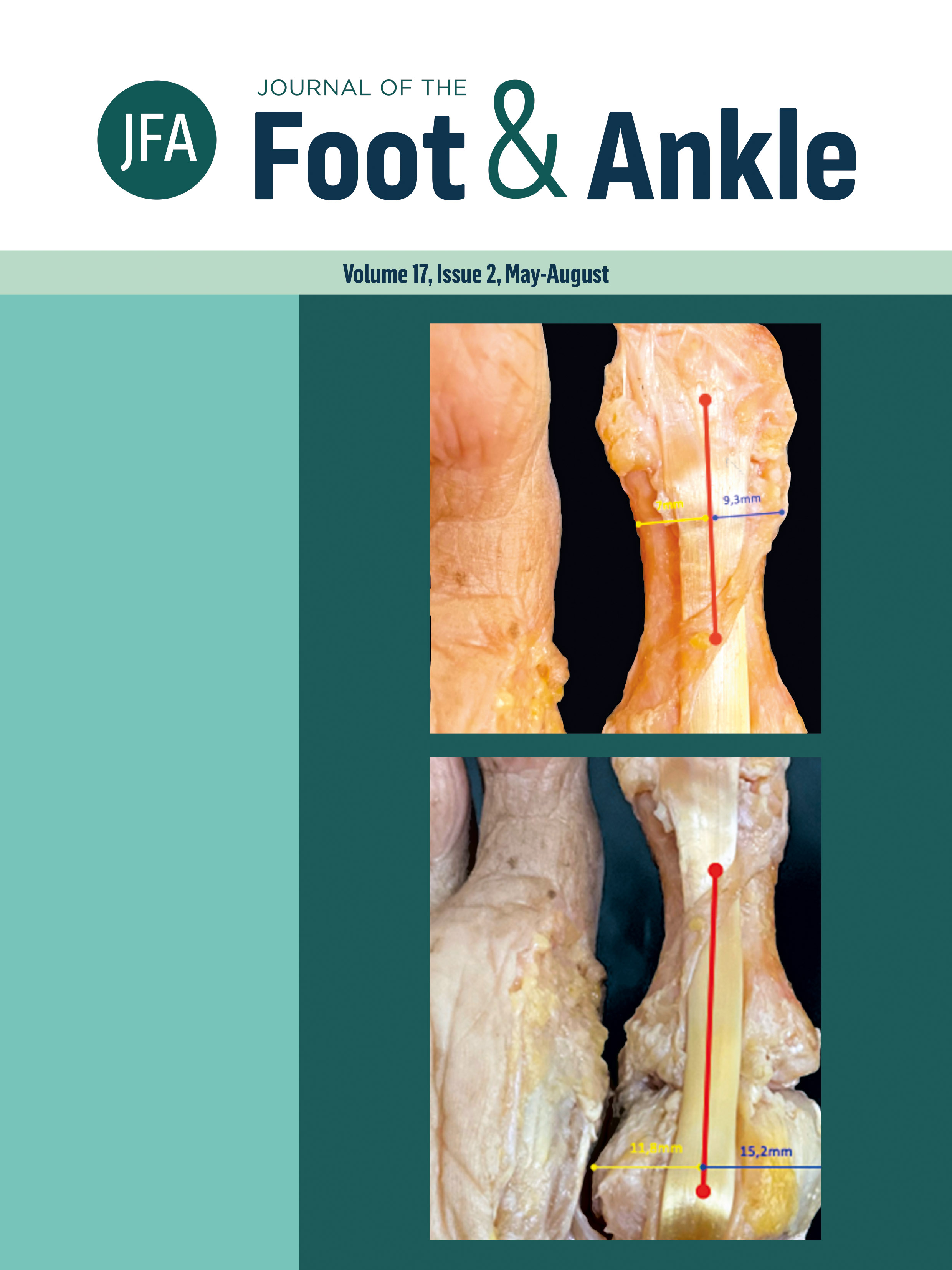
Methods: Ten cadaver feet were dissected to evaluate FHL distal insertion, the width of insertion, and the distance between insertion borders and medial-lateral phalangeal borders.
Results: All specimens showed a lateral tendon fascicle inserted more lateral and distal than the main insertion. The mean lateral and medial insertion distance to the phalangeal border was 3 mm and 5.2 mm. The FHL long axis was 12.36% laterally deviated at the metatarsophalangeal (MTP) joint and 14.07% at the interphalangeal (IP) joint.
Conclusion: The FHL has a closer insertion to the lateral phalanx border, and its long-axis midpoint is laterally located in relation to the IP and MTP joint. The detailed knowledge of the FHL true anatomy. The discovery of a lateral deviated axis, a lateral fascicle, and a lateral footprint.
Downloads
Published
How to Cite
Issue
Section
License
Copyright (c) 2023 Journal of the Foot & Ankle

This work is licensed under a Creative Commons Attribution-NonCommercial 4.0 International License.


