
Multimodal analysis and reconstruction of medical images in the evaluation of foot and ankle pathologies: Clinical and technological perspectives
DOI:
https://doi.org/10.30795/jfootankle.2026.v20.1915Keywords:
Foot; Ankle; 3D reconstruction; Weight-bearing; Computed tomography; Artificial intelligence; Augmented realityAbstract
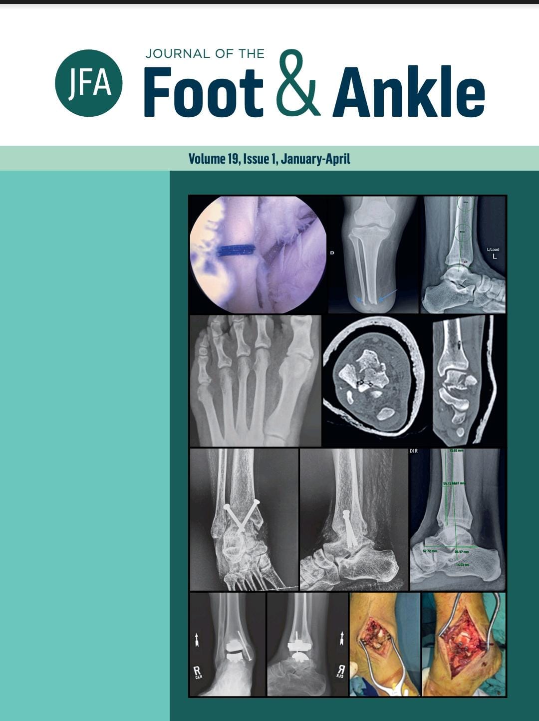
Objective: To analyze recent literature on 3D reconstruction technologies and multimodal fusion of medical images in orthopedics, with a focus on foot and ankle applications, and to discuss their potential, limitations, and future directions. Methods: This narrative review included original articles from 2010 to 2024 from PubMed, Scopus, and IEEE Xplore databases, using keywords related to “foot,” “ankle,” “3D reconstruction,” “weight-bearing computed tomography,” “machine learning,” “augmented reality,” and “multimodal fusion.” Studies on 3D reconstruction, image evaluation algorithms, and integration of imaging modalities in orthopedics were selected, with particular emphasis on those related to the foot and ankle. Those not affiliated with the medical field or specialty were excluded. The extracted data covered authorship, year, imaging modality, population, objective, and main conclusions Results: Twenty one studies were included in four categories: (1) standing weight-bearing computed tomography (WBCT) of the ankle and knee (4 studies), with greater precision and reproducibility than 2D radiographs; (2) 2D-3D algorithms (6 studies) based on neural networks and statistical models, capable of generating 3D models from conventional exams; (3) machine learning (3 studies) for fracture classification and ligament diagnosis, with high accuracy in automatic detection; and (4) augmented/mixed reality (8 studies) applied to surgical navigation and training, improving accuracy, reducing surgical time and radiation, in addition to showing educational potential. Conclusion: 3D reconstruction and multimodal fusion technologies provide new tools for evaluating foot and ankle pathologies. WBCT remains the gold standard for visualizing dynamic changes, but its restricted access can be mitigated using artificial intelligence for 3D reconstructions from conventional examinations. Advances in augmented reality and multimodal image fusion will permeate surgical diagnosis, planning, and execution, adding precision and safety in clinical practice. Level of evidence V; Expert opinion; Therapeutic studies - investigating the results of treatment
Downloads
Published
How to Cite
Issue
Section
License
Copyright (c) 2026 Journal of the Foot & Ankle

This work is licensed under a Creative Commons Attribution-NonCommercial 4.0 International License.


