Radiological evaluation of the longitudinal arch after posterior tibial tendon transfer
DOI:
https://doi.org/10.30795/2595-1459.2018.v1206Keywords:
Flatfoot, Tendon transfer, Peroneal nerveAbstract
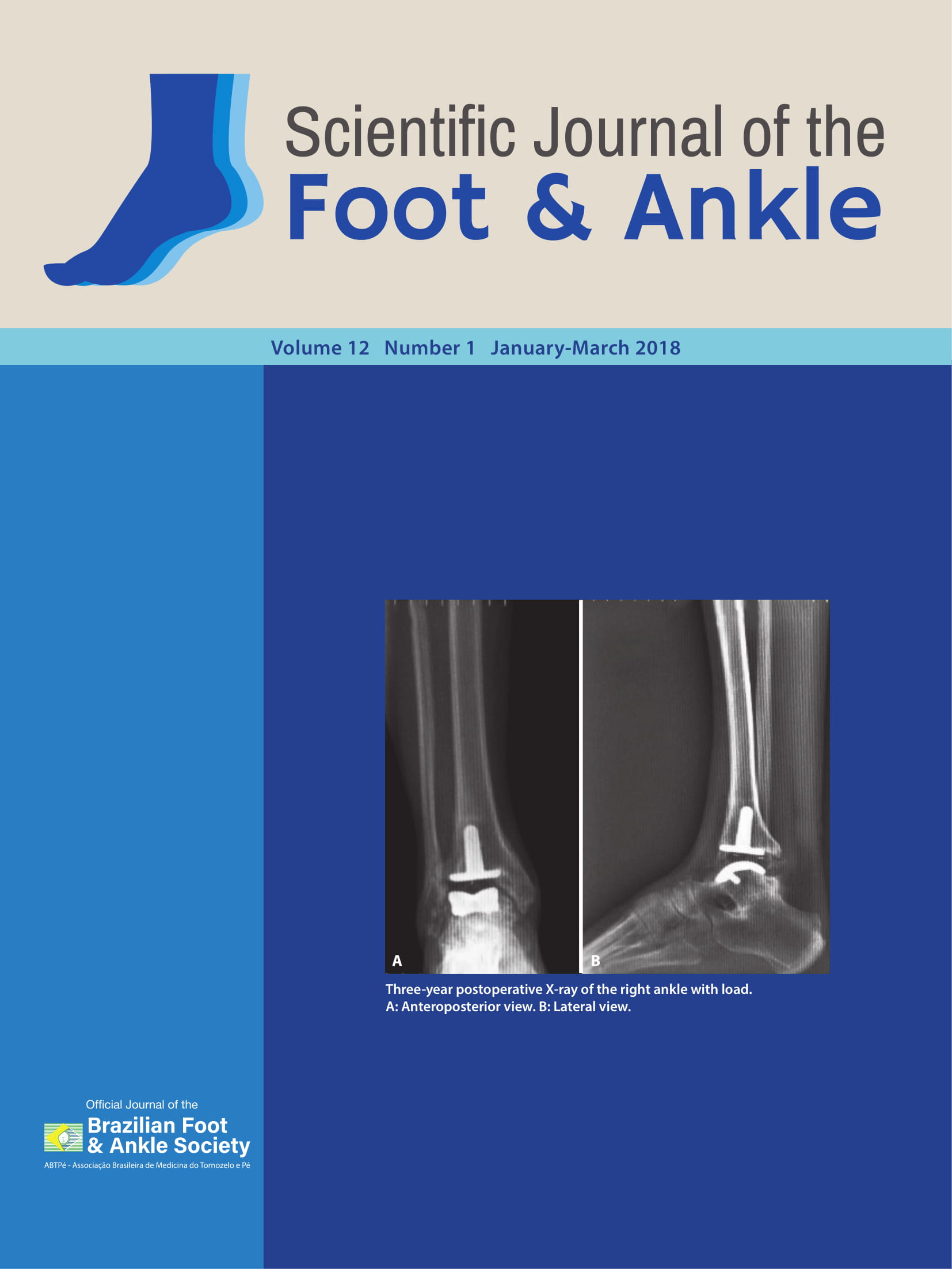
Objectives: To evaluate radiographic findings of medial longitudinal arch decreases of the foot based on examinations performed before and after posterior tibial transfer surgery to treat motor deficits caused by complete lesions of the peroneal nerve. Methods: A descriptive and analytical study was conducted based on information collected from medical records. Patients with at least two years of follow-up after surgery were included, and their radiographs were evaluated before and after the procedure. In the radiographs with the anteroposterior incidence of the foot, the talocalcaneal, talometatarsal and talonavicular congruence angles were evaluated. In the lateral view, the talocalcaneal, Meary’s and calcaneal pitch angles were analyzed. Data were collected regarding patient profiles, trauma mechanisms and follow-up times. Results: One patient had radiographic results suggestive of a decrease in the plantar arch after posterior tibial tendon transfer. Angular variation among the patients, which was within the normal range, was not significant. Conclusions: No significant decreases in the longitudinal arch of the foot were observed in the studied patients. Level of Evidence III; Retrospective Comparative Study.